The human brain is remarkable. In comparison to other animals, the brain is seven times bigger than it should be for the size of the body. During evolution, it enlarged in a rather short amount of time allowing humans to surpass other primates.
Researchers now have insight into the human advantage and ways to optimize the brain. We will explore these findings as well as the intricate functions of the nervous system.
The Human Advantage
The human advantage is not due to the size of the brain; it is the fact that humans have more neurons that carry and process information in a relatively small cerebral cortex (outer surface of the brain) than any other animal.
Thanks to the invention of cooking, humans were able to ingest a greater number of calories and nutrients in a short time. This lead to the rise in the human’s ability to reason, discover patterns, develop technology, and expand language.
The brain has approximately 86 billion neurons and is part of a complex, sophisticated system. The nervous system controls and coordinates all bodily functions and activities—every heartbeat, breath, movement, and thought.
- Whole Brain Size and General Mental Ability: A Review – an examination of the relationship between the brain size and mental ability, both within and between species
- The Elephant Brain in Numbers – a discussion of the superior cognitive abilities of the human brain in comparison to larger brains
- Equal Numbers of Neuronal and Nonneuronal Cells Make the Human Brain—an Isometrically Scaled-Up Primate Brain – an explanation of how the widespread quote that the human brain contains 100 billion neurons is found to be false
Cells of the Nervous System
Fresh neurons arise
Call it neurogenesis
New tricks for old brains
The Little Book of Neuroscience Haikus by Eric H. Chudler, PhD
For many years it was believed that nerve cells in the adult brain, once damaged or dead, did not replace themselves. Researchers, however, have found that neurogenesis, the growth and development of new neurons, is found in adults in several areas of the brain.
There are two broad categories of cells within the nervous system: neurons and glia.
- Neurons are known as the working units that process information and carry electrochemical impulses, which are electric and chemical messages. The chemical messages are known as neurotransmitters (e.g., serotonin, dopamine, and glutamate).
- Glial cells provide metabolic and mechanical support to the neurons as well as produce myelin (definition below).
The neuronal cell structure, or soma, includes a nucleus, cytoplasm, and cytoplasmic organelles. The nucleus contains deoxyribonucleic acid (DNA) and data that is necessary for growth, metabolism, and repair. Cytoplasm, also called protoplasm, is the colorless substance that fills the cell, which includes all the chemicals and parts necessary for the cell to function. The cytoplasmic organelles are known as “little organs” in the cytoplasm; each type of organelle has a specific role.

Figure 1: Neuron: Typical Structure. (© User: Ajimonthomas)/Wikimedia Commons/CC BY-SA 4.0)
The cell membrane surrounds each neuron and separates the contents of the cell from its surrounding environment. It controls what enters and leaves the cell and responds to signals from the environment.
Dendrites are the short, branched segments on the receiving end of the nerve cell.
The axon is the threadlike part of the nerve cell that extends from the cell body to meet and deliver impulses to another nerve cell. The length of an axon can range from a fraction of an inch to several feet.
Myelin is a fatty white sheath that surrounds the axon and some nerve cells. It forms an electrically insulating layer that is essential for the quick transmission of impulses along nerve cells.
Synapses are tiny gaps between neurons where chemical and electrical signals move from one neuron to another.
- Inside the Brain of a Neuron – a review of several studies to enhance the understanding of information processing in single-neurons arithmetic.
- Myelin and Nerve Structure – an illustration of the myelin sheath of a neuron.
- Glial Cells of the Peripheral Nervous System: Satellite Cell and Schwann Cell – a look into the glial cells of the peripheral nervous system.
- Regulation and Function of Adult Neurogenesis: From Genes to Cognition – a look into the relationship of neurogenesis and cognitive deficits in the adult human.
- Adult Neurogenesis in Humans: Common and Unique Traits in Mammals – a review of data on the extent and dynamics of neurogenesis in adult humans and other mammals.
- Brain-Derived Neurotrophic Factor (BDNF) and Its Clinical Implications – an explanation of the role BDNF plays in neuronal growth and survival.
- Action Potential for Kids – a description of resting membrane potential (neuron at rest) and action potential (neuron sending information down an axon).
- Neuroscience for Kids – a review of how to classify neurons based on the number of extensions that extend from the soma.
Organization and Functions of the Nervous System

Figure 2: Components of the Nervous System. (© Jenna Fair/Wikimedia Commons/CC-BY-3.0)
The Central Nervous System
The organs of the central nervous system include the brain and the spinal cord. Although recognized as two separate organs, the brain and spinal cord are continuous at the foramen magnum, which is a hole at the base of the skull forming the passage through which the spinal cord exits the cranial vault.
Because they are vital to human survival, they are encased in bone for protection. The brain sits in the cranial vault (the space within the skull), and the spinal cord is located in the vertebral column within the vertebral canal (spinal canal).
In addition to bone, meninges, or connective tissue, and cerebrospinal fluid surround and protect the membranes of the CNS. The three layers of meninges include the dura mater (outer), arachnoid (middle), and pia mater (innermost).
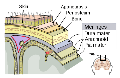
Figure 3: Meninges. (User: Mysid/Creative Commons/CC0 1.0)
Functions of the Brain
Nobody realizes that some people expend tremendous energy merely to be normal.
Albert Camus, Notebooks 1942-1951
Neurons need oxygen, glucose, and stimulation to function optimally. For example, if you have ever gone too long without eating, you may have felt sluggish from low blood glucose levels. The neurons in the brain have a high energy demand and consume approximately 5.6 mg of glucose per 100 g of human brain tissue per minute—approximately 25% of the body’s energy budget.
The brain divides into left and right cerebral hemispheres, which are joined by and communicated through the corpus callosum (figure 4).
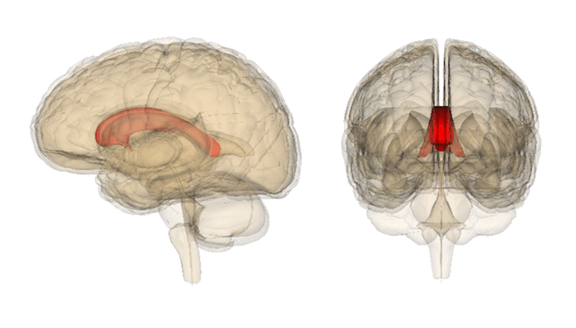
Figure 4: Corpus Callosum. (© User: Was a bee/Wikimedia Commons/CC BY-SA 2.1)
In neuroscience, the cerebrum and cerebral cortex are practically, but not precisely, the same thing.
There is a slight difference:
- The cerebrum is the entire top part of the brain including the white matter, which is mainly myelinated axons.
- The cerebral cortex technically refers to the highly wrinkled outer surface, or the gray matter, which contains numerous cell bodies and few myelinated axons.
The brain consists of three basic units: forebrain, midbrain, and hindbrain. Figure 5 illustrates each unit in a developing embryo.
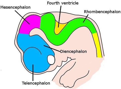
Figure 5: Six-Week Embryo Brain. (User: Kurzon/Wikimedia Commons/CC0 1.0)
Forebrain
The forebrain, also called prosencephalon, separates into the telencephalon and diencephalon, which are interconnected with the limbic system.
The telencephalon, or cerebrum, includes the cerebral cortex, which subdivides into four lobes (figure 6):
| Lobe | Basic Function |
|---|---|
| Frontal | Responsible for planning, reasoning, parts of speech, problem-solving, movement, and perception |
| Parietal | Integrates sensory information, which includes spatial orientation, movement, recognition, and perception of stimuli |
| Occipital | Involves visual processing |
| Temporal | Responsible for recognition and perception of auditory stimuli, speech, and memory |

Figure 6: Brain Lobes. (© BruceBlaus/Wikimedia Commons/CC BY-SA 3.0)
Parts of the diencephalon are as follows:
| Structure | Basic Function |
|---|---|
| Thalamus | Relays sensory and motor signals to the cerebral cortex, regulates consciousness, sleep, and alertness |
| Hypothalamus | Coordinates both the ANS and the activity of the pituitary—it controls temperature, hunger, thirst, sleep, and emotional and motivational behavior (the hypothalamus includes the *posterior pituitary gland*, which secretes oxytocin and vasopressin |
| Epithalamus | Links the nervous system to the endocrine system through the pituitary gland—it controls body temperature, thirst, hunger, fatigue, sleep, and circadian rhythm |
| Pineal body | Small endocrine gland that is a part of the epithalamus that produces melatonin to modulate sleep patterns, regulate new bone metabolism, and protect against neurodegeneration; it may also influence drug metabolism |
| Subthalamus | Plays a role in motor control and is interconnected with the basal ganglia (described below) |
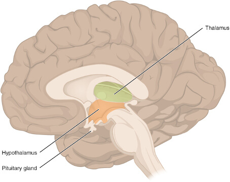
Figre 7: The Diencephalon. (© User: Openstax/Creative Commons/CC BY-SA 3.0)
Other major components located within and at the base of the cerebral cortex are as follows:
| Structure | Location | Basic Function |
|---|---|---|
| Cingulate cortex | Medial aspect of the cerebral cortex, above the corpus callosum | Responsible for emotion formation and processing, memory, learning, and links behavioral outcomes to motivation |
| Amygdala | Base of the cerebral cortex | Involved with memory, emotional reactions, and decision making |
| Hippocampus | Base of the cerebral cortex | Promotes short- and long-term memory, spatial memory, and is the center of mood and emotion—it is also a unique structure where new neurons can be generated |
| Olfactory bulb | Base of the cerebral cortex | Transmits smell information from the nose to the brain |
| Basal ganglia | Base of the cerebral cortex | Involves voluntary motor movement, procedural learning, and routine behaviors or habits (such as eye movements, teeth grinding, emotion, and cognition) |
Midbrain
The midbrain, or mesencephalon, is a section of the brainstem that forms the primary link between the forebrain and the hindbrain. It controls motor and visual functions, auditory responses, and sleep/wake arousal. It is made up of the tectum, tegmentum, cerebral aqueduct, and cerebral peduncles.
Hindbrain
The hindbrain, or rhombencephalon, occupies the posterior region of the brain and is a part of the brainstem. The hindbrain consists of the myelencephalon and metencephalon.
- The metencephalon portion of the hindbrain includes the cerebellum (Latin for “little brain”), pons, and reticular formation (located throughout the brainstem and cerebral cortex). The pons and the cerebellum are responsible for the control of movement, balance, and transmitting sensory information. The reticular formationis responsible for sleep and consciousness.
- The myelencephalon is also called the medulla oblongata, or the medulla. It deals with the vital autonomic functions of the body, such as digestion, respiration, and heart rate. Meditation may influence the medulla and the immune system.

Figure 8: The Brainstem. (© User: Openstax/Creative Commons/CC BY-SA 3.0)
Cranial Nerves
Twelve pairs of cranial nerves emerge directly from the brain; 10 of the 12 originate from the brainstem, and two nerves arise from the cerebrum. All of these nerves, except for the vagus nerve, pass through the foramina of the skull. A very small 13th cranial nerve, known as the terminal nerve, was identified more than a century ago; however, more research is needed to determine its role in humans.

Figure 9: The Cranial Nerves. (© User: CFCF/Wikimedia Commons/CC-BY-4.0)
| # | Name | Basic Function |
|---|---|---|
| I | Olfactory | Sense of smell |
| II | Optic | Sense of sight |
| III | Oculomotor | Controls motor movement of the eye and eyelid |
| IV | Trochlear | Controls an extraocular muscle that assists with visual tracking |
| V | Trigeminal | Responsible for motor innervation to muscles of mastication (chewing) and sensory innervation of the face |
| VI | Abducens | Eyeball movement |
| VII | Facial | Muscles of the face (facial expression) and taste |
| VIII | Vestibulocochlear | Hearing and balance (sense of body position) |
| IX | Glossopharyngeal | Swallowing and taste |
| X | Vagus | Gut (gastrointestinal tract), heart, and larynx |
| XI | Accessory | Sternocleidomastoid and trapezius muscles |
| XII | Hypoglossal | Tongue muscles |
Functions of the Spinal Cord
Downward from the medulla oblongata is the spinal cord, a long white tube that is made up of nervous tissue and support cells. The spinal cord divides into 31 segments with each giving rise to a pair of spinal nerves (peripheral nerves).
- 8 cervical (C) nerves – in the neck and upper spinal cord
- 12 thoracic (T) nerves – chest region
- 5 lumbar (L) nerves – middle to lower back
- 5 sacral (S) nerves – lower back
- 1 coccygeal (Cx) nerve – low back
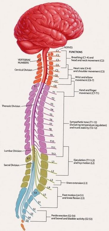
Figure 10: Anatomy of the Spine. (© University of Wisconsin School of Medicine and Public Health/No alterations made to image)
Three primary functions of the spinal cord include:
- Carrying information – The spinal cord sends signals to and from the brain.
- Coordinating reflexes – The spinal cord can coordinate reflexes on its own, which makes it responsible for quick, coordinated responses to stimuli. The thermal grill illusion is an excellent example this. It is an experiment that elicits an ice-like burning feeling—without causing harm—by using warm and cool metal bars. When a person places their hand on the metal bars, the pain signals quickly tell the spinal cord to react without knowing what it’s reacting to.
- Controlling reflexes – The spinal cord controls reflex responses that are typically involuntary. These reactions are sudden and are an automatic response.
The Peripheral Nervous System
The peripheral nervous system (PNS) consists of the nerves and ganglia. Nerves are bundles of nerve fibers, similar to how muscles are bundles of muscle fibers. Gangliaare clusters of nerves cells.
The spinal and cranial nerves extend from the CNS to peripheral organs. The basic constituents of the neural circuits within the PNS include afferent (sensory) neurons, efferent (motor) neurons, and interneurons (relay) neurons.
The PNS further subdivides into the somatic nervous system, autonomic nervous system, and the visceral nervous system. Further discussion will focus on the somatic and autonomic divisions of the peripheral nervous system.
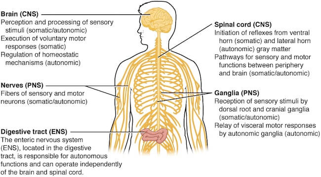
Figure 11: Somatic-Autonomic-Enteric Structures. (© User: CFCF/Wikimedia Commons/CC-BY-4.0)
Functions of the Somatic and Autonomic Nervous Systems
The somatic nervous system (SoNS), also called the voluntary nervous system, permits conscious control of the skeletal muscles and consists of both afferent nerves and efferent nerves. The SoNS includes spinal nerves, cranial nerves, and association nerves.
The autonomic nervous system (ANS), or involuntary nervous system, supplies motor impulses to the cardiac muscle, smooth muscle, and to the glandular epithelium (one or more cells that can produce and secrete a particular product). The ANS is only in connection with the efferent side of the nervous system.
The ANS further subdivides into the sympathetic, parasympathetic, and enteric nervous systems. The sympathetic and parasympathetic branches interact to maintain cardiovascular activity within an optimal range and permit reactions to changes in internal and external conditions.
- The sympathetic nervous system initiates the “fight-or-flight” mechanism when it detects danger or a threat. This threat may include internal stressors (e.g., chronic stress or anxiety) or external stressors (e.g., physical injury or environmental pollutant). A redirection of energy takes place when the sympathetic nervous system is activated; digestion is put on hold, the heart rate and breathing rate escalate, pupils dilate, and there is an increase in the production of saliva and sweat.
- When the parasympathetic nervous system takes over, the opposite effect occurs; digestion restarts and breathing and pupils return to normal.
- The enteric nervous system, also known as the intrinsic nervous system, is a separate nervous system for the gastrointestinal system, which to a great extent, autonomously controls bowel motility. The gut-brain axis (GBA) consists of a bidirectional connection between the enteric and central nervous systems. The GBA links cognitive and emotional centers of the brain with peripheral intestinal functions. Recent advances in research describe the importance of the gut microbiotain influencing these interactions.
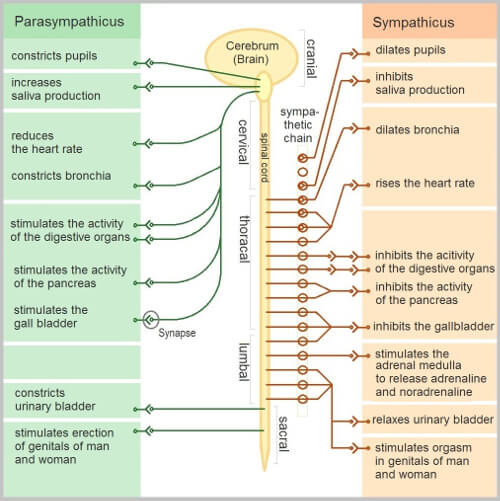
Figure 12: The Autonomic Nervous System. (User: Geo-Science-International/Wikimedia Commons/CC0 1.0)
Signs and Symptoms of Nervous System Disorders
The brain gives clues about its state of health. The following are the most common key indicators of a nervous system disorder; however, each person may experience symptoms differently.
- A headache that changes or is different or persistent
- Blood pressure changes
- Slurred speech
- Sudden loss of sight or double vision
- Impaired mental ability
- Memory loss
- Lack of coordination
- Muscle rigidity and wasting
- Weakness or loss of muscle strength
- Loss of feeling or tingling
- Back pain that radiates to the feet or another part of the body
- Tremors and seizures
- A nervous system disorder may resemble other medical conditions or problems
Disorders of the Nervous System
If not quickly and appropriately treated, a disease or illness that involves the nervous system may have severe consequences. As you review the list below you will find that many disorders fit into several categories.
Nerve and Blood Flow Disruption (Blood Supply to the Brain: Video Tutorial)
- Transient ischemic attack (TIA)
- Stroke
- Epidural hematoma
- Subdural hematoma
- Subarachnoid hemorrhage
- Low Blood Pressure
- Anemia
Structural Disorders
- Carpal tunnel syndrome
- Childhood tumors in the brain or spinal cord
- Adult central nervous system tumors
- Meningiomas
- Injury to the brain or spinal cord
- Cervical spondylosis
- Peripheral neuropathy
- Bell’s palsy
- Trigeminal neuralgia
Functional Difficulties, Degeneration, and Autoimmune
- Alzheimer’s disease
- Epilepsy
- Multiple sclerosis
- Huntington’s disease
- Parkinson’s disease
- Amyotrophic lateral sclerosis (ALS)
- Narcolepsy
- Guillain-Barre syndrome
Infections
Optimize Your Brain and Prevent Injury
By following the guidelines below, people can help keep their nervous system healthy. Always speak to a healthcare practitioner before starting any new exercise, regime, or diet.
- Exercise regularly—get your heart rate up to increase BDNF.
- Get plenty of rest and sleep.
- Refrain from caffeine six hours before bedtime.
- Take care of conditions that may cause a decrease in the function of the nervous system, such as low or high blood pressure and diabetes.
- Eat a balanced diet that consists of whole food with ample sources of flavonoids, omega-3 fatty acids, B6, B12, and folate.
- Get plenty of sunshine and have your vitamin D checked.
- Ask your doctor if nootropics are right for you.
- Perform intermittent fasting or caloric restriction.
- Improve your gut-brain axis and intestinal motility: video tutorial.
- Talk to your doctor about a low-carbohydrate diet for mood disorders or an autoimmune diet.
- Drink plenty of water—the heart and brain are composed of approximately 73% water.
- Stop or limit alcohol.
- Do not smoke or use tobacco products.
- Do not use illegal drugs such as meth, cocaine , heroin, or other non-prescribed drugs (such as non-prescribed opioids).
- Learn new skills to rewire the brain.
- Concentrate on one thing at a time—the brain is not the best multitasker.
- Wear head protection when performing a risky activity, which may include riding a bike, skateboarding, football, etc.
- Prevent falls in the home—remove rugs, keep walkways clear, clean up clutter, and use a nightlight.
Please contact us at support@pacificmedicaltraining.com to reach the author or recommend other ailments or brain-health tips that interest you.
 Written by Sarah Gehrke, MSN, RN and last updated Nov 5, 2017
 Last reviewed by Michael A. Tomeo MD on Nov 5, 2017